NU4 PET IQ
Brand: Leeds Test Objects | Category: Nuclear Medicine & Imaging
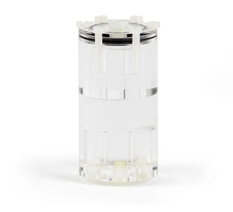
A small field of view PET image quality phantom designed for pre-clinical scanners. It allows testing of image quality, attenuation correction and scatter correction using fillable rods, fillable cylinders and a uniform section, to the NEMA NU4 standard.
Plan your specification
Match NU4 PET IQ to the research scanner and test
Leeds documents NU4 PET IQ for small-field PET image quality and correction tests, citing NEMA NU4-2008. Confirm the intended measurement and configuration in the purchase specification.
- Geometry and positioning
- The published outer cylinder diameter is 33.5 mm. Check the scanner's usable field of view and holder clearance, and ask whether the required positioning accessories are included.
- Protocol and analysis
- Name the test and standard edition your team will use. Ask the responsible physicist to identify any additional test objects, software or instruments; this phantom alone does not demonstrate completion of every scanner performance test.
- Supplied components and handling documents
- Request the current specification, instructions, closures and replacement-part details. Confirm preparation and source requirements through the qualified research team rather than assuming radioactive material is part of the supply.
Product documentation
Technical Specifications
| Parameter | Value |
|---|---|
| Cylinder diameter | 33.5 mm |
| Specification | 5 fillable rods, 1 to 5 mm diameter |
| Specification | 2 fillable cylinders of 8 mm diameter, separated from the main chamber by a 1 mm wall |
| Specification | 1 uniform fillable section, 15 mm thick |
| Specification | Conforms to NEMA NU4-2008 |
Get a Quote
Request an itemised quotation covering the proposed configuration, availability, lead time and agreed supply and support scope.
Useful details to share
- Scanner make/model and usable field of view
- Intended image-quality or correction measurements and protocol edition
Share what you know. You can start with your name and a phone number or email; final specifications can follow.
Not sure which configuration you need? Help me choose
Contact Vvon Technologies Limited about Leeds Test Objects equipment requirements in Bangladesh.
Phone: +8801805987890 | Email: info@vvon.com.bd
Back to Nuclear Medicine & Imaging
More from Nuclear Medicine QA Phantoms
- PET CT MRI Cubes
A set of seven clear PMMA cubes each holding a small fillable spherical void, used as point source markers for checking alignment between PET, CT and MR. They can be filled with a radionuclide, CT contrast agent or MR contrast agent and sealed with the nylon screw supplied.
- MicroPET PSF
A tissue equivalent point spread function phantom for pre-clinical PET systems. It comprises pairs of cylinders in six tissue equivalent materials, one solid and one with a central hole to take a capillary filled with radionuclide.
- PET IQ
An anthropomorphic PMMA body phantom for PET and PET/CT image quality testing. Six hollow fillable spheres screw into a plate on one face and can be filled with water for cold lesion imaging or with 18F FDG for hot lesion imaging.